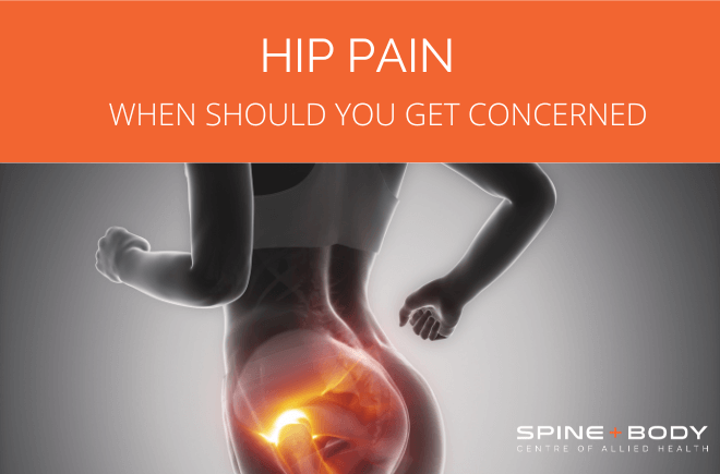
Hip pain – when should you get concerned?

By Peter Georgilopoulos, APA Sports Physiotherapist, Spine + Body Gold Coast.
We may all know of someone within our inner group that has undergone a Total Hip Replacement [THR] procedure and may feel intuitively that this is a common procedure, which, relatively speaking, it is and becoming increasingly more so. Despite the increasing prevalence of elective joint replacement surgery, the true incidence as a representation of our population is far less than we perceive it to be however. If we evaluate statistical data of hip replacement procedures in the United States and draw parallels to similar per capita numbers in Australia, we would expect around 0.83% of the population to undergo such a procedure within our community and approximately the same throughout the Western World. In essence, roughly speaking, this represents less than one person per hundred so if we were to estimate that we have social contact with, say, 500 people on a regular basis, we could reasonably expect there to be five or so people that we know that would be functioning without pain courtesy of a prosthetic hip joint. What we also know is that women represent marginally higher numbers than men and numbers increase with age reaching 5.26% of the population by 80 years of age. There are a number of issues that may explain the reason for greater representation of women compared to men- one might be the greater Q angle [the angle of the femur or thigh bone in relation to the pelvis in a wider female pelvis] and possibly the greater incidence of osteoporosis [de-mineralisation of the bone cortex due to hormonal changes] amongst women. Perhaps because of the relatively common nature of this procedure, many people that develop a gradual onset of hip pain immediately fear that they are imminently due for joint replacement surgery which in the majority of cases is far from the truth. Needless to say, many people self- diagnose and avoid seeking professional advice believing that they will be steered towards hip replacement surgery well before any diagnosis is made.
Where is the pain located
It is important to categorically state that the majority of self-diagnosed hip ailments are not in fact related to the hip joint at all but may fall outside the parameters of the joint itself. Most of these, of course can be treated rapidly and effectively with minimally or entirely non-invasive techniques and procedures.
If the presenting pain is located to the outside of the pelvis, specifically if one can pin point an area of focal pain just above the joint, the likely diagnosis is “ trochanteric bursitis” [inflammation of the bursa ]. All joints have a bursa [or bursae in plural] which creates the intra- articular fluid that “lubricates” the cartilaginous surface and provides nutrient input to the articular surface of the joint. Excess amount of joint fluid is created whenever the joint is placed under immense load or trauma and this can occur through a direct blow to the region or as a result of repetitive overload attributable to poor biomechanics such as a tight psoas major which is a powerful muscle connecting the lumbar spine to the hip joint. Often low back pain leads to activation of this muscle creating excessive internal rotation at the hip joint with every step, leading to joint inflammation and distension of the hip bursa. Stretching or releasing the psoas muscle, improving hamstring flexibility and hip joint “turnout” [to use a ballet term to describe the ability to externally rotate the hip] will inevitably improve the biomechanics and reduce symptoms. Anti- inflammatory medication and / or localised cortisone injections can also assist in rapidly easing the inflammatory symptoms.
If pain is localised to the buttocks or towards the back of the pelvis, the cause of pain may be neural irritation [such as low grade sciatic pain especially if pain increases in sitting] or may represent a degenerative tear of one of the stabilising gluteal muscles which is often an indication once again of excessive internal rotation at the hip joint.
What signs may indicate some internal derangement within the hip joint proper?
The first is that pain may occur predominately on weight bearing although at the later stages of hip joint degenerative changes, the hip joint surface may be exposed to such a degree that there may be constant pain present. It is important to note that any internal hip degenerative condition will invariably refer pain to the adductor or groin area towards the pubic region. An easy clinical test that can be self- administered to evaluate whether symptoms are stemming from some intrinsic hip joint lesion or some extraneous inflammatory soft tissue issue is the quadrant position. The “quadrant” position of any joint is the specific position in which the two joint surfaces are in greatest contact with each other. In this position, any pathology within the joint surface is most likely to be provoked consequently reproducing “the pain”. The quadrant position for the hip joint is examined with the patient lying on their back. The hip and knee joint is bent to 90 degrees, the leg is then pushed towards the opposite leg and finally the hip joint itself is rotated internally. The combination of all three movements brings the joint surfaces of the hip joint into greatest opposition or compression exposing any internal lesion as “groin” pain.
So what does this mean and what should happen next?
Firstly, a positive quadrant test warrants further investigation of the hip joint surface as there may be a number of causes for the presenting symptoms. The most obvious cause may be significant osteoarthritis or degenerative changes involving the joint surface but this may not always be the case. Other considerations may be a labral tear within the joint surface. The labrum is a cartilaginous disc similar to that found within the knee whose function is to dampen load bearing forces between the joint surfaces. A split or tear may occur in sudden or forced hip movements such as a slip on a wet floor leading to “ the splits” or stepping into a pot hole resulting in sudden jarring or commonly whilst water or snow skiing when the legs come apart suddenly. More often than not this requires arthroscopic debridement [trimming] to avoid further joint erosion. An MRI or CT scan can assist in specifically identifying such a lesion.
Other causes
Other causes of hip joint erosion can be an impingement issue which is commonly described as a CAM or Pincer deformity. These descriptive terms relate to developmental anomalies within the neck of the femur or to the outer rim of the “socket” or acetabulum of the joint. A straight Xray can confirm the presence of any such lesion as these are bony configurations which are easily viewed. Early surgical intervention will stave off further degenerative changes which can also lead to a labral tear as a further complication.
In some cases undiagnosed congenital hip dysplasia [displacement of the hip within its socket at birth] results in malformation of the joint surface leading to early degenerative changes. In these cases specialist evaluation is strongly recommended. Perthes disease is also a condition that creates malformation and a broadening of the articular surfaces which inevitably leads to true hip pain. This also requires specialist care in the early stages of diagnosis.
Osteoarthritis
Above all, the most common issue with true hip joint pain is due to osteoarthritis. If symptoms are unilateral, look for some degree of biomechanical asymmetry such as leg length discrepancy or significant structural scoliosis as a likely cause. Falls in the elderly are also a common cause of possible fractures of the neck of the femur which can be internally fixed but in most cases require a Total Hip Replacement procedure as the flow of blood to the head of the femur may be disrupted leading to avascular necrosis. Failure of any prosthetic joint appliance usually occurs between one or two decades and it is not, as is commonly believed, due to failure of the prosthetic component but rather a dislodging of the interface between the prosthesis and living tissue [bone]. Invariably this occurs as the bony cells are replaced in time so that the bond created by the connecting cement no longer exists resulting in loosening of the prosthesis. When this occurs revision surgery is required so timing of any prosthetic surgical option is vital.
Having stated all of the above, it is important to reiterate that for the most part the majority of “hip “ issues are extraneous to the hip joint proper and generally respond quite rapidly to specific medical and other rehabilitative measures.
Book your appointment with Peter Georgilopoulos at Spine + Body.
Peter is Director and Founder of Spine + Body Centre of Allied Health on the Gold Coast ph: 07 5531 6422 or contact us by email here.


