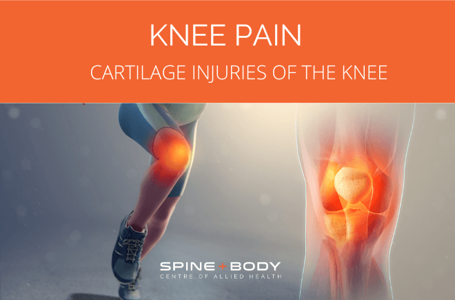
Cartilage injuries of the knee

By Peter Georgilopoulos, APA Sports Physiotherapist, Spine + Body Gold Coast.
Most people might be surprised to learn that the most common injury incurred by breast- stroke swimmers is not to the upper limb as one might expect but rather to the inside aspect of the knees. Many may also be surprised to discover that knee injuries occur more frequently in netball than in Australian Rules or either of the Rugby codes.
There are obviously many structures situated in and around the knee that can be damaged resulting in pain and dysfunction but there is one in particular that generally occurs as a consequence of what might appear to be an innocuous incident. A typical history might be getting out of a low car by placing one leg out of the vehicle and transferring body weight onto the leading leg during the process of standing up. Another seemingly innocuous mechanism of injury might be squatting down to retrieve an item out of arm’s reach under a bed. In both incidents, there may be little in the way of pain or swelling to suggest that a significant knee injury has occurred. The injured subject might be perplexed by the onset of swelling, pain and an inability to weight bear normally the following day. Some may even experience a locked knee in which they might be unable to bend or straighten the joint. Any attempt to straighten a locked knee results in severe pain causing further confusion and distress.
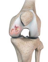
The heading of this article offers a not- so cryptic clue as to the likely cause of injury. Confusing the issue is the term “cartilage” which is commonly used to describe two distinct anatomical structures within the joint. Medically, the term “cartilage” describes the shiny, “porcelain-like” covering protecting bones within a joint. Colloquially however, the term “cartilage” is used to describe what is medically referred to as a “meniscus”. This is a crescent shaped structure situated between the two bones of the knee [the tibia or “shin-bone” and the femur or “thigh bone”]. The meniscus is detached for the most part except for two fixation points on the tibial surface [tibial plateau] at the front and back known as the anterior and posterior horn respectively. There are two menisci in each knee [medial and lateral or “inside” and “outside”] giving the appearance of two crescent shaped structures facing each other if one could view the middle of the knee joint from above.
Their function is to act as “spacers” between the two weight bearing bones, protecting the joint surface from erosion and enhancing joint compliance [the ability for two surfaces to fit together]. The menisci consist of cartilaginous material, hence the colloquial reference. It is very conceivable that trauma may occur to either the cartilaginous joint surface, one or other meniscus, or both so adhering to accurate descriptive terms may be far more useful in describing the injury medically although making that distinction may not be as critical in every -day life.
There are numerous descriptive terms that identify various meniscal lesions:
- A “bucket- handle” tear describes a peripheral lesion which remains at
 tached at either end. This can become problematic if the fragment detaches from the alignment of the main meniscal body creating a mechanical obstruction to free movement within the joint. In some cases, the flap may return to a less provocative position spontaneously partially restoring movement and reducing pain.
 - A Parrot Beak lesion describes a wedge shaped split from the periphery to the inner body of the meniscus.
- Other terms such as “horizontal”, “peripheral” and “displaced” are self -descriptive.
It is important to establish how far the lesion extends within the body of the meniscus which may determine the likelihood of gradual healing and repair or the need for surgery. The fundamental issue is that the outer third of the meniscus has a blood supply, albeit a very poor one, so lesions within this zone can reasonably be expected to repair over time if protected. The inner two- thirds however are avascular [there is no blood supply to this zone] so there is no ability to create healing scar tissue. The greatest consideration as to whether a peripheral tear is likely to heal depends on the alignment of the fragment to the remaining meniscal body and the avoidance of rotation weight-bearing movements that are likely to disrupt the repairing structure.
Surgical options [arthroscopic debridement or “trimming”] may be recommended as a result of either clinical assessment or radiological confirmation but the primary objective must be to preserve meniscal integrity where possible. Obviously, surgically extracting some of the meniscus will precipitate possible earlier degenerative changes within the exposed joint surface over time but then again so would persisting with a dislodged fragment that continues to traumatise previously unaffected portions of the joint surface. The decision of whether to proceed to surgery or persist with protected repair over time is unique to each case. The one self- determining presentation is in the case of a locked knee which almost inevitably results in surgery. An aside to surgical debridement of a torn meniscus is that typically a surgeon will bevel the outer margin of the torn fragment so as not to present a sharp edge where the fragment has been removed but this obviously thins the leading edge of the newly repaired structure, potentially elevating the possibility of further future tears. To optimise surgical outcomes, surgeons are increasingly preferring to suture [stitch] the fragment when this presents itself within the outer vascular third. The benefits are restoring as much of the meniscus as possible and therefore minimising future adverse outcomes but the negatives are that the time to healing is considerably longer and requires more extensive rehabilitation under specific weight bearing protocols.
Anatomical and postural variations of the knee may significantly predispose some to meniscal tears. Knocked knee [Genu Valgum] or Bow Legged [Genu Varum ] postures asymmetrically load either the inner or outer compartment thereby scuffing the meniscal smooth surface increasing the likelihood of a tear as a result of any forceful rotational action. Without question, the most provocative action that leads to meniscal tears is twisting or rotation on bent knee and this is precisely why dancing, football , breast-stroke swimming, netball and gymnastics [amongst others] present a higher risk of injury.
Clinical examination by a trained medical or allied health professional is essential in the early stages. X-rays are not useful diagnostic tools in this regard as menisci are not radio- opaque [not visible]. An MRI or CT scan is far preferable as both enable visualisation of meniscal structures as well as any other ligamentous or capsular structures that may be implicated. When meniscal damage is suspected, crutches are recommended to avoid further trauma, ice can be used for 10-15 minutes daily to minimise swelling and above all, joint rotation is to be avoided, especially when getting in and out of a car.
Book your appointment with Peter Georgilopoulos at Spine + Body.
Peter is Director and Founder of Spine + Body Centre of Allied Health on the Gold Coast ph: 07 5531 6422 or contact us by email here.


